Looking inside ephemeral ultrabright x-ray laser pulses
November 09, 2021NUS scientists co-lead an international collaboration to resolve the elusive wavefronts of x-ray free-electron lasers, paving the way towards high-throughput, high-resolution, machine-learning-enabled imaging.
Bright, ultrafast X-ray Free-electron Laser (XFEL) pulsed sources can interrogate millions of single particles in mere hours. By imaging each particle separately, XFELs open up an unprecedented window to subtle but important differences in each particles’ structure and dynamics. However, this form of single-particle imaging comes with a formidable machine learning task: each particle introduces several unmeasured nuisance parameters that must be inferred (particles’ orientation, position, state, etc); compounded on these are differences between different XFEL pulses’ intensity and wavefront profiles. These pulses are notoriously difficult to characterise at the focus, because they are so intense that they easily obliterate conventional instruments used to characterise x-rays.
A research team lead by Prof Duane LOH from both the Department of Biological Sciences and Department of Physics, National University of Singapore in collaboration with international partners comprising groups from Sweden, United Kingdom, Italy and United States of America were able to use a technique known as mixed-state ptychography to resolve many thousands of XFEL pulses from the Linac Coherent Light Source at the SLAC National Accelerator Laboratory. Unlike conventional visible light-based microscopy, which uses physical lenses to form images, the structure of XFEL-illuminated particles must instead be inferred using computational lenses (such as ptychography) that form images using known principles of x-ray-based science and optics. The additional complication is that no two XFEL pulses are alike due to the way they are spontaneously generated. The mixed-state formalism handles this uncertainty remarkably well. Their technique has the additional advantage of using very little signal strength. This is important because the power of the x-ray pulses had to be highly attenuated to protect the illuminated reference target.
The key differences between thousands of x-ray pulses were quantitatively captured and shaped into important priors for structure determination and experimental design.
Prof Loh said, “XFEL-based imaging experiments can sometimes feel like playing several chess games at once in a blindfolded state. Our work finally reveals which imaging parameters are critical for resolving the structural classes hiding within the particle ensemble.”
This work demonstrates a drop-in scheme to rapidly characterise and optimise tens of thousands of extremely bright and focused x-ray pulses within minutes. The eventual goal is to do the same while imaging — sipping away a small fraction of unscattered x-ray photons from each pulse to perform truly live and concurrent diagnostics.

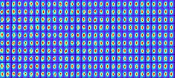
Random variations in the intensity profiles of hundreds of focused x-ray free-electron laser pulses (shown here) reduces imaging resolution. The authors of this work combined machine learning with computational lenses to infer and reduce these variations, thus improving single-particle imaging resolution.
References
[1] Daurer BJ; Sala S; Hantke MF; Reddy HKN; Bielecki J; Shen Z; Nettelblad C; Svenda M; Ekeberg T; Carini GA; Hart P; Osipov T; Aquila A; Loh ND*; Maia FRNC; Thibault P, “Ptychographic wavefront characterization for single-particle imaging at x-ray lasers” OPTICA Volume: 8 Issue: 4 Page: 551-562 DOI: 10.1364/OPTICA.416655 Published: 2021.
[2] Cho DH; Shen Z; Ihm Y; Wi DH; Jung C; Nam D; Kim S; Park SY; Kim KS; Sung D; Lee H; Shin JY; Hwang J; Lee SY; Lee SY; Han SW; Noh DY; Loh ND*; Song CY*, “High-Throughput 3D Ensemble Characterization of Individual Core-Shell Nanoparticles with X-ray Free Electron Laser Single-Particle Imaging” ACS NANO Volume: 15 Issue: 3 Page: 4066-4076 DOI:10.1021/acsnano.0c07961 Published: 2021.
[3] Ayyer K*; et. al., “3D diffractive imaging of nanoparticle ensembles using an x-ray laser” OPTICA Volume: 8 Issue: 1 Page: 15-23 DOI: 10.1364/OPTICA.410851 Published: 2021.


