New radiobiology beamline at CIBA
December 11, 201711 Dec 2017. NUS physicists in collaboration with clinicians from the National Cancer Centre Singapore (NCCS) are developing a new beamline to advance proton therapy in cancer treatment.
Radiotherapy is a technique that is now used widely for the treatment of cancer. Conventional radiation therapy utilises energetic photons (e.g. x-rays) to target cancerous tumours in the body. As the photon beam intensity decreases exponentially when it penetrates through biological matter, the positioning and dosage of the beam need to be very precisely controlled to minimise damage to healthy tissues. Protons interact with matter differently. Most of the energy dissipation from the protons happen in the vicinity where the particle stops. The velocity of the initial beam of protons can therefore be adjusted such that they slow down, upon passing through tissue, and stop at different depths within the body. This means that protons can be directed to “damage” the tumour while leaving surrounding healthy tissues relatively unaffected.
Prof Andrew BETTIOL from the Department of Physics, NUS is constructing a new beamline at the accelerator facility in the Centre for Ion Beam Applications (CIBA) located at NUS. This beamline is part of a research programme which brings together scientists from NUS and clinicians from NCCS to study the fundamental effects of proton irradiation on various types of cancerous and non-cancerous cells for cancer treatment.
Prof Bettiol said, “When it is ready, this new radiobiology beamline will have the unique capability to target sub-cellular structures in live cells with energetic protons. The beamline has a confocal microscope that provides the user with real-time visualisation of the cellular response to a controlled irradiation dose. The proton beams can be focused to as small as 300 nm in diameter. This is 1,000 times smaller than the width of a human hair. Such a small beam size allows us to target sub-cellular structure with a precise amount of radiation.”
The beamline is expected to be completed by the first quarter of 2018.
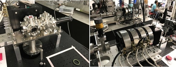
Figure 1 shows photos of the new radiobiology beamline in CIBA (still under construction).
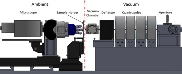
Figure 2 shows a schematic drawing of the beamline system. The beamline will have a set of magnetic quadrupole lenses to focus the proton beams down to approximately 100 nm. The protons emerge from the vacuum system through a custom designed nozzle that can be placed in close proximity to a cell culture dish. The cells can be individually targeted with a controlled irradiation dose of protons and the changes viewed in real-time using a microscope.
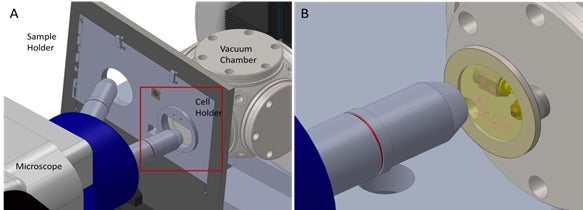
Figure 3 shows a close-up schematic of (A) the cell holder and (B) the imaging system.