Next generation fast proton imaging and fabrication
Jeroen Anton VAN KAN ((Group Leader, Physics) ) March 22, 201722 Mar 2017. NUS physicists have designed and successively micro-fabricated a miniature ion beam source prototype, paving the way to improve ion beam brightness by up to a million times.
| Microscopy has been an integral part of scientific development. Miniaturisation has spurred development in many fields. Fast proton microscopy has several advantages over traditional forms of microscopy. A fast incoming proton interacts mainly with substrate electrons. Due to the mass mismatch between protons and electrons, a proton beam follows almost a straight path through the material. In proton-electron collisions, the substrate electrons get just enough energy to break connecting bonds within a range of about 1 nanometre(nm). Current proton microscopes are typically very large in size and not user-friendly. However, the main weakness in a proton microscope is the ion source, which is typically several million times less in brightness compared to other competitive beam sources.
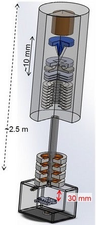
Recent experimental tests by Prof Jeroen Anton VAN KAN and his collaborators with “Lab on chip ion sources” have shown breakthrough results, opening up the way to improve ion beam brightness by up to a million times! [1,2] This will have major implications. Firstly, it potentially allows the spot size for fast protons to breach the single digit nm regime. Secondly, it allows the possibility of miniaturising the size of a proton microscope to that of a table top system. Developing a compact proton microscope will democratise the ion beam community, making it accessible to a much wider audience. Prof van Kan is now researching the development of a table-top proton microscope system [3]. Next generation proton microscopes can find applications in many diverse areas. Proton beam writing (PBW) is able to achieve a much better resolution compared to electron beam writing, aiming for sub-nm details without having “proximity effects” (the final pattern obtained being wider than the scanned pattern, due to interactions of the electron beam with the material), a major drawback in electron beam writing systems. By using different ion species, it can potentially be used for fabricating next generation quantum computers. In the biomedical field, proton microscopy allows for sub 10 nm whole cell imaging. This will open up new pathways to investigate and understand the absorption of nanoparticles by cells in the development of better drug delivery methods. Protons with energies at or below 0.5 MeV will cause double-strand breaks in DNA. This result can potentially be used to improve cancer treatment in radio biology using fast protons. This research is supported by research grants from the Singapore Ministry of Education Tier 1, the Singapore National Research Foundation Competitive Research Programme and the US Air Force. |

Figure shows a compact proton beam writing system. |
References
1. Liu N; Xu X; Pang R; Raman PS; Khursheed A; van Kan JA*, “Brightness measurement of an electron impact gas ion source for proton beam writing applications” REVIEW OF SCIENTIFIC INSTRUMENTS Volume: 87 Issue: 2 Article Number: 02A903 DOI: 10.1063/1.4932005 Published: 2016
2. Xu X; Pang R; Raman PS; Mariappan R; Khursheed A; van Kan JA*, “Fabrication and development of high brightness nano-aperture ion source” MICROELECTRONIC ENGINEERING Volume: 174 Pages: 20-23 DOI: 10.1016/j.mee.2016.12.009 Published: 2017
3. Xu X; Liu N; Raman PS; Qureshi S; Pang R; Khursheed A; van Kan JA*, “Design considerations for a compact proton beam writing system aiming for fast sub-10 nm direct write lithography” NUCLEAR INSTRUMENTS AND METHODS IN PHYSICS RESEARCH SECTION B: BEAM INTERACTIONS WITH MATERIALS AND ATOMS DOI: 10.1016/j.nimb.2016.12.031


