Super-resolution imaging using MeV ion beams
Andrew BETTIOL ((Group Leader, Physics) ) December 07, 20167 Dec 2016. NUS physicists have developed a new form of super-resolution fluorescence microscopy for biomedical applications by using highly focused mega-electron volt(MeV) ion beams.
Optical super-resolution microscopy through the use of nonlinear properties of fluorophores, structured illumination or the stochastic nature of light emission, has emerged in recent years as an important technique for imaging sub-cellular structure. An alternative method of achieving super-resolutions is through the use of charged particles such as electrons or ions. Charged particles have much smaller de Broglie wavelengths. This means that diffraction effects are absent at nanometre spatial dimensions, allowing imaging of structures beyond optical resolutions. The main limitation of particle based microscopy is the penetration depth. When imaging thick biological samples such as whole cells (up to 10 microns thick), high-resolution images can only be obtained of the surface of the sample.
A research team led by Prof Andrew BETTIOL from the Centre for Ion Beam Applications (CIBA) in the Department of Physics, NUS has constructed a system for simultaneous fluorescence and structural imaging of whole cells by using a beam of highly focused (MeV) ions. MeV ions are able to penetrate many tens of microns into biological samples without any degradation of the image resolution. The new system has achieved spatial resolutions down to sub-30 nanometres. This is more than 10 times below the optical diffraction limit. The fluorescent probes used for labelling the cells were developed from upconversion nanoparticles (NaYF4:Yb/Tm) which had been optimised for their brightness and resistance to degradation under ion beam irradiation.
The combined fluorescence and structural imaging capability allows for the quantification of fluorescent nanoparticles in whole cells. This methodology is expected to pioneer the development of more advanced imaging applications at the subcellular level. An example of such an application could be the quantitative measurement of intracellular bio-distribution of drugs delivered by fluorescent nanoparticles.
Future work will focus on the functionalisation, targeting and ion beam imaging of nanoparticles used for probing important biological and biomedical processes.
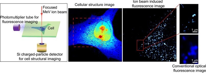
The figure demonstrates fluorescence and structural imaging of a whole HeLa cell by using a beam of MeV helium ions focused to a 20-nanometre spot size. The region marked by a red-dotted square in the cellular structure image was imaged in ion beam induced fluorescence. Comparison between the ion beam induced fluorescence image and a conventional optical fluorescence image shows the much improved spatial resolution that can be obtained by using MeV ion beams.
Reference
Mi ZH; Zhang YH; Vanga SK; Chen CB; Tan HQ; Watt F*; Liu XG*; Bettiol AA*, “Subwavelength imaging through ion-beam-induced upconversion” NATURE COMMUNICATIONS Volume: 6 Article Number: 8832 DOI: 10.1038/ncomms9832