Tortuous path to nanocrystals
Utkur MIRSAIDOV and Duane LOH ((Group Leaders, Physics and Biological Sciences) ) November 21, 201621 Nov 2016. NUS researchers observed the crystallisation of gold particles in real time in an aqueous solution in clear violation of classical nucleation theory.
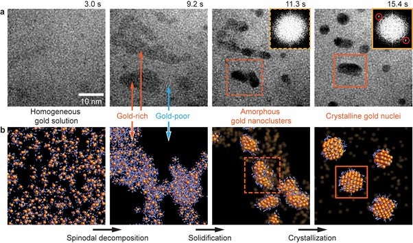
Crystallisation is ubiquitous in many natural and man-made processes, yet our knowledge of it is still very limited. A multi-disciplinary team of researchers from NUS, the Agency for Science Technology and Research and the University of Chicago, co-led by Prof Utkur MIRSAIDOV and Prof Duane LOH, both from the Department of Physics and Department of Biological Sciences, NUS have revealed the surprisingly complex and elusive steps in crystallising nanocrystals from solution. They showed that crystals emerge from a solute-rich solution in three steps (see figure): first, the solution spontaneously separates into solute-poor and solute-rich regions; structurally amorphous nanoclusters form within the solute-rich regions; then these nanoclusters reorganise into nanocrystals from which larger crystals grow.
Despite decades of research, scientists still cannot reliably predict if, when, and how nature assembles an ordered crystal from a dynamic and violent storm of disordered atoms. The common wisdom is that crystals emerge from nanometre -sized versions of themselves known as nuclei that are thought to form spontaneously. Understanding how nuclei form, via a process known as nucleation, is key to explaining why crystallisation often occurs unexpectedly. Ideally, we would want to watch how single nuclei emerge fromasolution. However, tracking pre-nucleation steps has been challenging since nucleation occurs randomly, deep within a solution, at nanometre scales, and only very briefly.
The research team has now developed a new method for illuminating the mysterious intermediate steps leading to crystallisation in various systems. While there were prior hints of these intermediate states in other systems, this work reports the first direct observation of how these intermediate states are related and unfold in real time. It conclusively shows that “spinodal decomposition”, a process from which solute-rich phases can spontaneously appear in otherwise homogeneous solutions, acts as a pre-step to crystal nucleation. Understanding this decomposition may one day help to control nucleation in natural or artificial systems.
At the heart of this discovery are the quantitative electron microscopy techniques and microfabrication processes developed at the Centre for Bio-imaging Sciences and Centre for Advanced 2D Materials at NUS that enable probing of real-time dynamics within very thin liquid films. These techniques allow the researchers to discern how crystal nuclei form in multiple steps, transitioning between intermediate states before finally reconfiguring into nano-crystals. The challenge now is to capture and understand the collective dynamics of the atoms that trigger each step of nucleation. Together with a number of recent works1, it is clear that electron microscopy is rapidly moving from static images to dynamic movies. The work reported here shows how quantitation and statistics have become integral to understanding stochastic and heterogeneous dynamics at the atomic scale. Moving forward, data science will play an increasing role in expanding the capabilities of electron microscopy to image messy dynamics in nature.

Figure shows the three-step pathway for gold nucleation in a solution.(a) Frames from a Transmission Electron Microscope movie showing the intermediate steps in nucleating gold nanocrystals. Solution spontaneously de-mixes into gold-poor and gold-rich liquid phases (lighter and darker regions, respectively). Amorphous gold nanoclusters emerge from gold-rich phases that then crystallise into crystalline nuclei. Crystallinity of nuclei is revealed by Fourier Transform showing the gold {111} face-centred cubic reciprocal lattice spacings circled in red. Insets show Fourier transforms of cropped square regions reveal that crystallisation with spacing circled in red. (b) Schematic depiction of nucleation pathway (gold atoms and nearby water molecules are coloured in orange and blue, respectively).
For more details of their work, please see:
www.mirsaidov.org
www.duaneloh.com
Footnote 1: Panciera, F. et al. Nature Mater. 14, 820–825 (2015); Liao, et al. Science 345, 916-919 (2014).
Reference
Loh ND; Sen S; Bosman M; Tan SF; Zhong J; Nijhuis CA*; Kral P*; Matsudaira P; Mirsaidov UM*, “Multistep nucleation of nanocrystals in aqueous solution”, NATURE CHEMISTRY doi:10.1038/nchem.2618


