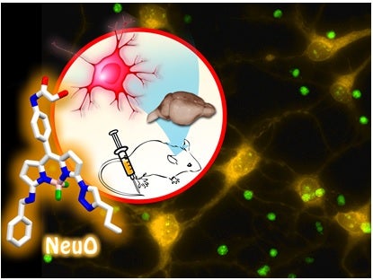
Recently, a team led by Prof CHANG Young-Tae from the Department of Chemistry discovered NeuO, which is a simple chemical dye that can selectively label neurons fluorescently over other brain cells. After further optimization, NeuO can also be applied for generic imaging of neurons in the whole body of small animals like mouse and zebrafish.
NeuO can be a valuable tool for illuminating the intricate neuronal networks that govern neural function and to visualize how neurons differentiate, develop dendrites synapses and interact with other cells in the brain. Furthermore, due to NeuO’s selectivity in only staining live neurons, the probe can also be potentially utilized to enlighten us on how various neurological disorders, such as Alzheimer’s and Parkinson’s disease (the primary target being neuron degeneration), progress.
For decades, scientists have been searching in vain for such a chemical tool that can label live neurons. The simple chemical dyes that have been developed are generally unselective or work only for dead neurons. Fluorescent proteins, although successful, are not so straightforward to use and more importantly, have very limited opportunity to be developed for clinical use. The discovery of NeuO fulfills this unmet need for a simple and reliable tool to illuminate live neurons performing its natural function.

NeuO fluorescently labels live neurons selectively in vitro, ex vivo and in vivo. [Image credit: ER Jun Cheng]
Reference
Er JC, Leong C, Teoh CL, Yuan Q, Merchant P, Dunn M, Sulzer D, Sames D, Bhinge A, Kim D, Kim SM, Yoon MH, Stanton LW, Je SH, Yun SW, Chang YT. “NeuO: a Fluorescent Chemical Probe for Live Neuron Labeling.” Angew Chem Int. Ed 54 (2015) 2442


