Diffraction before destruction
Duane LOH (Group Leader, Biological Sciences) () April 26, 201626 Apr 2016 Scientists in NUS have demonstrated how x-ray lasers could help us image biological macromolecules in water.
Nanometer-sized biological molecules are difficult to resolve because they are fragile: they come apart when they are imaged with energetic x-rays or electrons. Fortunately, ultra-short x-ray laser pulses billions of times brighter than previous x-ray sources can now illuminate single biomolecules to produce faint but meaningful signals. These signals are then statistically combined to yield structural information. Because these x-ray pulses are so short, they flee from biomolecules before the latter get a chance to move or show damage. This property permits so-called “diffraction before destruction”, where scientists can image unsuspecting and unperturbed nanoscale objects in their native environment [1,2].
For decades, people have imaged nanoscale biomolecules by coercing many of their copies into a crystal, because millions of such crystallised molecules in unison can scatter enough x-rays to make their structures visible. Many biological macromolecules, however, do not naturally crystallise to facilitate crystallography. The “single-particle imaging” paradigm with pulsed x-ray lasers aims to resolve previously unseen proteins one at a time, in their native environments even if they do not crystallise or are mutually dissimilar. The task of assembling structural information from these heterogeneous measurements is then borne by computation and statistical means.
Hundreds of experimental parameters modify how x-ray free-electron lasers are generated, focused, made to interact with biomolecules, then detected, and analysed. The combinatorial complexity of such imaging experiments is staggering, which makes designing such experiments tricky, tedious and frustratingly uncertain. Dr Duane LOH from the Department of Biological Sciences in NUS, together with four other groups of scientists from Center for Free-electron Laser, DESY, Hamburg, Germany and the European XFEL GmbH, Hamburg Germany has created a comprehensive multi-physics framework that realistically simulates for the first time, how specimen damage in single-particle imaging can be mitigated by instrument and algorithm design [3].
Scientists can now quickly and affordably diagnose instrument issues using this framework, and test novel but otherwise costly schemes proposed to improve imaging conditions. In the next phase of this international collaboration, they will identify which are the most critical design parameters for single-particle imaging, and develop mitigation strategies where needed.

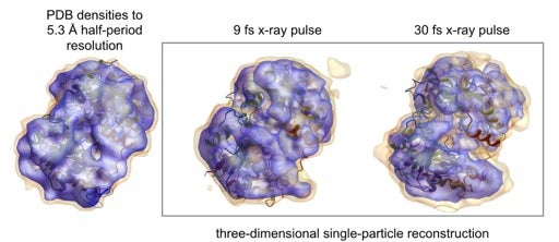
Figure shows (left) two-level contour plot of the ideal electron density map of a nitrogenase iron protein. (Box on the right) shows how these contours in reconstructed maps suffer with longer x-ray pulses [3]. Each map is created from hundreds of thousands of proteins, each damaged slightly differently and scattering only about 100 x-ray photons. Shorter pulses “see” less damaged proteins, but they produce less signals to learn the proteins’ structures. A multi-physics framework was created to tackle tricky questions such as “how much shorter can we make these x-ray pulses and still have enough signals left over for structure determination?” [Image credit: Duane Loh].
References
1. Loh ND, et al. “Fractal morphology, imaging and mass spectrometry of single aerosol particles in flight”. Nature 486 (2012) 513.
2. Ekeberg T, et al. “Three-Dimensional Reconstruction of the Giant Mimivirus Particle with an X-Ray Free-Electron Laser.” Phys. Rev. Lett. 114(2015) 098102.
3. Yoon CH, et al. “A comprehensive simulation framework for imaging single particles and biomolecules at the European X-ray Free-electron Laser.” Scientific Reports. 6 (2016) 24791. doi: 10.1038/srep24791.


